Mechanical forces in growing gut tissue shape villi in embryonic animals
The human intestine is a marvel of morphology, meandering 21 feet inside the abdomen, with a miniature forest of finger-like projections called villi covering its inner surface. Now a team including L. Mahadevan, Ph.D., a Wyss Institute Founding Core Faculty Member, has uncovered the forces that direct that forest to develop.
Mahadevan, who is also the Lola England de Valpine Professor of Applied Mathematics, a Professor of Organismic and Evolutionary Biology, and Professor of Physics at Harvard, uses the tools of applied mathematics, physics, engineering, and biology to uncover how matter is shaped, how it flows – and how its shape and flow are controlled: How does a cucumber tendril coil and wind? How do objects get stuck on a sticky surface? How do soap films form and deform?
More recently, Mahadevan, who goes by “Maha,” has investigated the forces that shape the gut. In 2011, he and his collaborators reported in Nature that the small intestine forms loops, much as a piece of paper curls when part of it is moistened. It and the tissue that anchors it grow at different rates, which cause the growing gut to curve.

Now, in the next stage of that collaboration, which includes Clifford J. Tabin, Ph.D., a Professor of Genetics at Harvard Medical School, Amy Shyer, Ph.D., a Research Fellow in Tabin’s lab, and Toumas Tallinen, a former postdoctoral fellow in Maha’s lab, the team has found a similar principle at work as the embryonic gut shapes its forest of villi. They reported their results in the August 29 issue of Science.
The results shed new light on gut development and raise questions about how mechanical forces on tissues regulate growth — and how they might go disastrously awry to cause cancer.
“Differential growth is a simple and principled way to pattern growing tissues, and it could underlie development of a variety of organs,” Maha said.
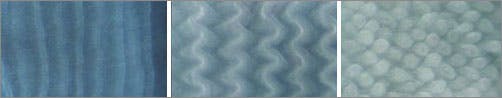
The gut’s villi are needed to help the organ take up nutrients more efficiently, and they develop early. To find out which forces induce them to develop, Maha and his collaborators removed the intestine of embryonic chickens at various stages of development. Then they fixed the tissue, treated it with dyes to help see various tissue types, and examined it under a microscope. They saw that villi form in three stages: First, the gut lining, which is initially smooth, develops ridges that run parallel to the gut itself. Those ridges then buckle into zigzags that remain flat on the gut lining. Finally, the elbow of each zigzag rises to form a bump that grows into a villus.
To find out whether mechanical forces triggered these changes, the researchers surgically teased apart layers of the embryonic gut wall. Without a particular muscle that surrounds the gut like a sausage casing, the gut tube grew fatter and failed to form ridges. Drugs that blocked muscle development led to the same result, as did encasing the widening gut in a tight silk casing. These results suggested that this muscle surrounds the gut, squeezing the growing inner layers of the gut wall until they buckle to form ridges.
To see what causes the ridges to buckle into zigzags, the researchers treated the gut with the same muscle-blocking drugs after they had formed ridges. This blocked a second layer of muscle that restrains the gut from growing longer, which kept the ridges from forming zigzags. The same drugs also kept the zigzags from popping up and forming villi. The results showed that mechanical forces drove all three stages of villi development.
To find out which mechanical forces underlay the third and final stage of villi development, the researchers rolled clay into long, thin threads that resembled an intestine, then bent it into zigzags and twisted it. The elbows of the zigzags rose into bumps that resembled budding villi in embryonic chicks.
To confirm that their model explained how villi developed, the team then built a mathematical and computational model of gut development. Using data on the dimensions and elasticity of embryonic gut’s tissue layers, the model predicted the full course of villi development. This was true for embryonic chicks — and for animals such as frogs, snakes and mice whose villi develop differently, or not at all.
Like all good science, the work raises as many questions as it answers, Maha said. Do mechanical forces induce biochemical or genetic changes in tissue that spur them to grow? Can too much or too little force on tissue remove the brakes on tissue growth, leading to cancer? Do mechanical forces direct development in other tissues besides the gut?
Only more science will answer these questions.