T cells, a subtype of white blood cells, play key roles in cell-mediated immunity, be it to fight infections and cancer or, when corrupted, to react against the body’s own cells in more than 80 autoimmune diseases, including type I diabetes, multiple sclerosis, rheumatoid arthritis and others. However, isolating disease-related T cells from the body to better study or eliminate them poses a formidable challenge to researchers and clinicians.
Wyss Institute researchers are generating implantable and injectable biomaterials to concentrate and trap disease-related T cells. To this end, and for a limited period of time, they place porous scaffolds with biological components under the skin. These spiked scaffolds can attract and ‘trap’ circulating T cells that after retrieval of the biomaterial can be easily isolated. Using this approach, researchers can get access to disease specific autoreactive T cells to study and better understand their detrimental functions. They can even deploy their T cell traps to potentially reduce the number of tissue-damaging T cells therapeutically, or ask how drugs affect disease-specific T cell populations in patients.
These T cell traps could give us a handle on autoreactive T cell populations in different autoimmune diseases and be used as a read-out for the drugs that may actually raise immune tolerance in these disorders.
The Wyss team provided proof-of-concept for the method by applying it to animal models of human type I diabetes and isolating diabetes-promoting T cell populations.
Wyss researchers are also expanding the use of T cell traps to capture T cells with pivotal roles in other than autoimmune diseases which will enable them to study their functions or exploit them therapeutically.

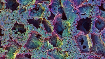
1/3 This scanning electron microscopy image shows a cross section of the porous biomaterial that can be implanted under the skin of patients to trap disease-causing T cells. Credit: James Weaver 
2/3 This scanning electron microscope image shows the T cell-trapping biomaterial up-close with cavities and ducts allowing the entry, as well as movement and binding of autoimmune T cells in its interior. Credit: James Weaver 
3/3 This is a histological section of retrieved beta-cell scaffolds stained for cellular structures 14 days after implantation. In the magnified area on the right, trapped autoimmune T cells are showing up as dark dots. Credit: Wyss Institute at Harvard University